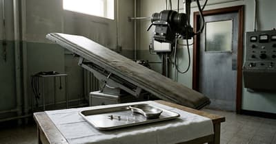
In the mid 1960s, neurosurgeon Ron Pawl watched a spinal X-ray test make the patient part of the machine. Pantopaque went into the spinal fluid. Then the table tipped head up or head down, and gravity pulled the oil through the canal while fluoroscopy watched where it went.[1]
Before CT and MRI made spinal imaging routine, doctors used Pantopaque, an oil-based dye that could show the spinal canal on X-rays but often could not be fully removed afterward.
On the fluoroscopy table, Pantopaque moved because the table moved. Pawl wrote that the medium was denser than water and loaded with iodine, which made it block X-rays and outline the soft tissue inside the spinal canal. To steer it, the staff used the table and the patient's body like a slow track for a bright bead of oil.[1]
After the images were taken, the bead still had to come back out. Pantopaque was not absorbable, Pawl wrote, so doctors tried to remove it by aspiration. The job was formidable. Some of the oil was often left behind, and repeat studies could show arachnoiditis, inflammation involving the membranes around the brain and spinal cord.[1]
Pantopaque, also called iophendylate or Myodil, belonged to the years when seeing the spinal canal meant moving an object through it. A 2022 Cureus report says doctors used it for myelography, ventriculography, and cisternography from 1944 until 1988. Water-soluble contrast agents, CT, and MRI eventually pushed it aside. The same report lists complications that helped end its run, including arachnoiditis, chronic irritation, and anaphylaxis.[2]
Half a century later, radiologists were still meeting the old oil. A 2023 case report described iodinated contrast deposits found 50 years after a previous myelogram, and noted that doctors had used aspiration cannulas to remove oil-based contrast after the procedure.[3] In a 2024 case, a 93-year-old man had a head CT after a fall, and the bright droplets on the scan were traced back to remote myelography rather than a new bleed.[4]
A modern scan can turn those droplets into a small diagnostic riddle. The 2022 report warned that Pantopaque remnants have been mistaken for tumors or shotgun pellets.[2] A test built for an older way of seeing the spine can still announce itself as a tiny white dot, long after the room, the table, and the person who held the syringe are gone.
Sources
- Ron Pawl, "History of myelography with pantopaque contributing to arachnoiditis," Surgical Neurology International
- Frank Ma et al., "Pantopaque: Does It Still Migrate With Gravity After 30 Years?," Cureus
- Imad-eddine Sahri et al., "Intracranial iodinated contrast medium deposits 50 years following a previous myelography," Radiology Case Reports
- Tom Saliba et al., "Subarachnoid Intra-cranial Lipiodol Deposits," Cureus